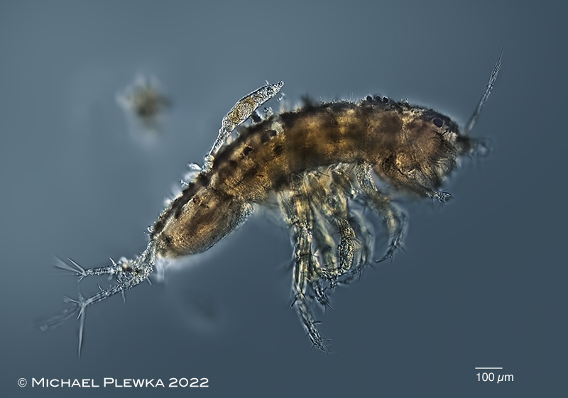
| Rotaria magna-calcarata; specimen on the dorsal side of the carapax of an amphipod crustacean (Gammarus sp.). (4) |
| |
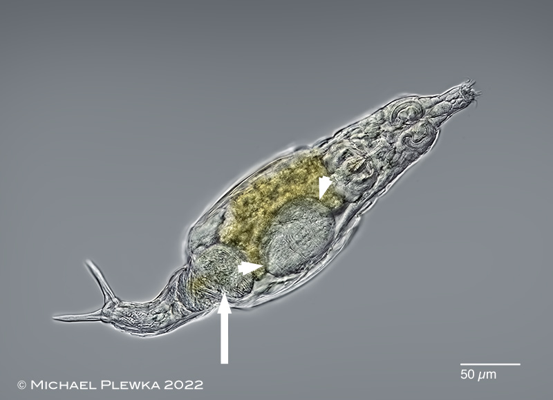 |
| Rotaria magna-calcarata; the arrow points to the bladder of this specimen which is infested by the parasite Leptoclava parasita. Also an embryo is visible (marked by arrowheads). (4) |
| |
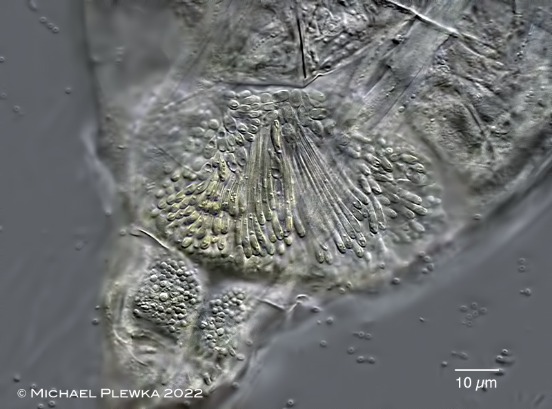 |
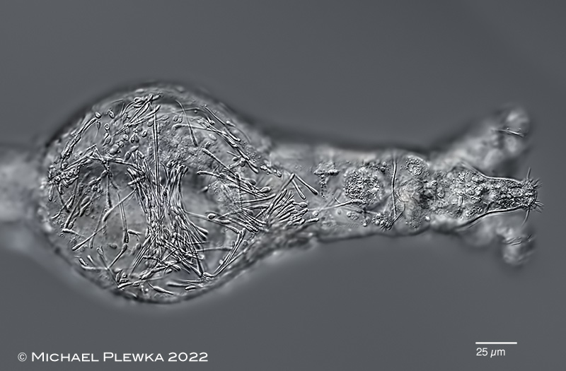
| Rotaria magna-calcarata; bladder of the above specimen is infested by the parasite Leptoclava parasita. L. parasita seems to infest mainly Rotaria species: also Rotaria macrura has been observed with this parasite. (4) |
| |
 |
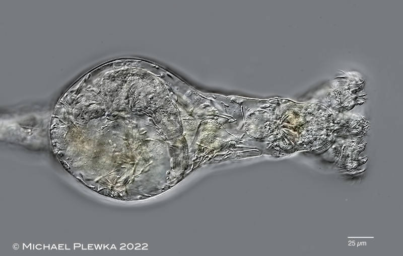
| Rotaria magna-calcarata; the above specimen was kept on a slide for several days and monitored every day. After 5 days the specimen did not move any longer, the corona was retracted; the rotifer seemed to be dead. At that time the parasites were distributed within the entire animal. When the water layer between coverslide and rotifer was reduced the trochal columns protruded and started whirling, but the rest of the specimen kept motionless except for the embryo that was also visible. It may be concluded that the movement of the trochal cilia is not triggered directly by neural activity but only by the extrusion of the trochal discs. |
| |
 |
 |
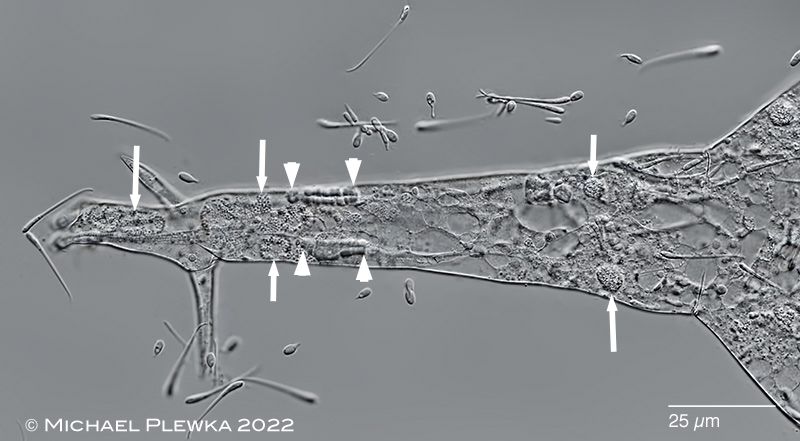
Rotaria magna-calcarata; two aspects of the anterior part. Upper image: crop of the above image. The parasites have infested the entire animal. While the trochal and rostral cilia were still beating the trophi did not move. Lower image: focus plane on the daughter rotifer which was still moving (except the trophi, which is not typical). Some of the other inner organs of the mother rotifer are partly destroyed: the longitudinal retractor muscles have desintegrated, also the stomach lumen is not visible (instead there is a "bubble"). Only in a single nephridial cell movement of the cilia was visible in the whole animal (when a (bdelloid) rotifer dies the nephridial cells are the last ones to stop their function). Because it would be normal that the mother dies only after giving birth to the daughter it is likely that these injuries of the mother is the result of the parasites.
Unfortunately it was not possible to isolate the daughter from the mother rotifer. As far as observation was possible, in the daughter, however, no parasites could be observed. |
| |
 |
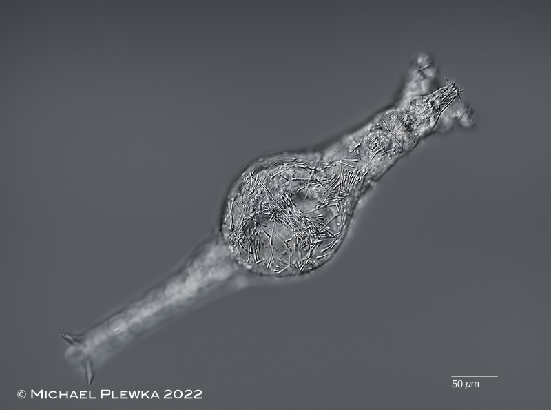
| Rotaria magna-calcarata; foot of slightly compressed specimen. The longitudinal retractor muscles have desintegrated, the remnants of the footglands are visible (arrowheads). The arrows point to some vesicles filled with moving particles (BROWN-movement?) which may be a different developmental stage of the parasite. |
| |
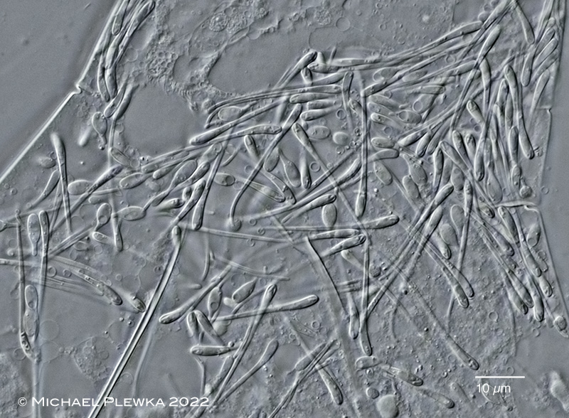 |
| Rotaria magna-calcarata; compressed specimen; Leptoclava-parasites |
| |
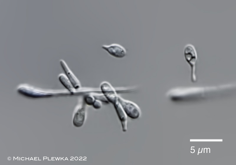 |
| Rotaria magna-calcarata; some other strange bodies, which may be other developmental stages of the parasite. |
| |
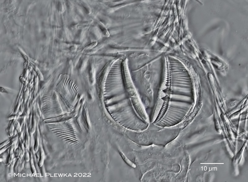 |
| Rotaria magna-calcarata; trophi of daughter (left) and mother (right) after maceration with SDS. When NaOCl was applied the trophi of the daughter dissolved, but the Leptoclava-parasites resisted this chemical treatment (and the mother trophi of course as well ) |
| |
| |
| |
|
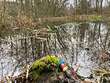
Location (3); (4): Sprockhoevel-Schee, NRW, Germany; pond. |
 |
| |
| Habitat (3); (4): plankton-sample with detritus (click image to enlarge >>>) |
| |
| Date (3): 27.05.2021 (3); 23.07.2022 (4) |
| |
|
|
|
|
|
| |
| |
|
|
|